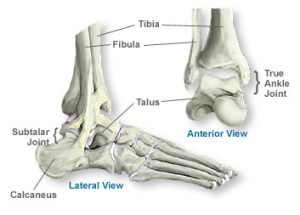
Foot Anatomy
Within the feet lay bones, joints, ligaments, tendons, muscles, nerves, and blood vessels. When discussing the anatomy of the foot, physicians divide the foot into three main parts; the forefoot, the midfoot, and the hindfoot.
Forefoot
The metatarsels, phalanges, and sesamoids make up the forefoot. Within the forefoot lay twenty one bones that break down into five metatarsels, fourteen phalanges, and two sesamoids. Each toe has a proximal, middle, and distal phalange with the exception of the big toe. The big toe only has a proximal and distal phalanx. The two sesamoid bones on each foot lay side by side, proximal to the proximal phalanx of the big toe. These sesamoid bones lay within the flexor hallucis brevis tendons. The metatarsels join the midfoot at the tarsal-metatarsal joint.
Midfoot
When doctors refer to the midfoot, they refer to the area of the foot starting at the transverse tarsal joint and ending at the tarsometatarsal joint. The navicular, cuboid, and medal, middle, and lateral cuneiforms make up the midfoot. The cuboid lays on the outside (lateral side) of the foot and articulates with the calcaneus bone to form the calcaneocuboid joint. The navicular lays distal to the talus and acts in conjunction with the talus to create the talonavicular joint. In addition to the talus, the navicular also connects with all three cuneiform bones. The three cuneiform bones aid in the high stability of the foot.
Hindfoot
Spanning from the ankle joint to the transverse tarsal joint, the hindfoot consists of the talus and the calcaneus. The calcaneus remains the largest bone in the foot and people commonly refer to the calcaneus using the term “heel bone”. The calcaneus helps form two joints. The calcaneus and talus form the subtalar joint while the calcaneus and cuboid bone form the calcaneal-cuboid joint.
The talus sits on top of the calcaneus to form the subtalar joint. The talus also helps form the talonavicular joint. Surrounded by hyaline cartilage, the talus allows for smooth movement of the foot. This hyaline cartilage makes it difficult to have a strong blood supply resulting in long healing times for talus injuries.
Muscles of the foot
The muscles of the foot work to move the foot and the toes of the foot. The foot can invert and evert. The toes can flex, extend, abduct (move away from the body) and adduct (move back toward the body).
Common injuries to the foot
The feet must support all the weight of the human body. Since the feet must support the weight in addition to have flexibility in order to walk, the feet find themselves susceptible to injuries. Injuries to the feet may occur do to traumatic injury like falling, dropping things onto the feet, or sporting events. Injuries to the feet may also occur due to age and wear and tear.
Common injuries and conditions of the feet include:
To view a list of all insurances that AOA Orthopedic Specialists accept, click HERE. To schedule an appointment online, click HERE.
EXPERIENCING foot PAIN? CALL 817-375-5200 TO SCHEDULE AN APPOINTMENT WITH AN AOA ORTHOPEDIC SPECIALIST TODAY!
FAQ
What is Plantar Fasciitis?
The plantar fascia is a thick band of tissue that runs along the bottom of the foot from the heel to the toes. Plantar fasciitis is a frequent condition that causes discomfort and inflammation in this region. Heel pain and stiffness are frequent symptoms, particularly in the morning or after extended periods of sitting or standing.
What are Bunions?
Bony lumps called bunions develop at the base of the big toe joint. An imbalance in the foot mechanics often brings them on, and wearing tight or narrow shoes may worsen them. Bunions may result in foot deformities, discomfort, and edema.
How can I keep my feet healthy?
It is crucial to wear comfortable, well-fitting shoes, maintain good foot cleanliness, and engage in regular foot exercises and stretches if you want to keep your feet healthy. Injuries and conditions of the foot can be decreased by maintaining a healthy weight and avoiding high-impact activities. Talk to an orthopedic surgeon in Dallas for more information.


