Parts of the Ankle; Ankle Bone Anatomy
The ankle joint, or Talocrural joint, is a large synovial joint. It is a hinge joint that allows plantarflexion and dorsiflexion, moving the foot up and down. The ankle is more stable while joint is in dorsiflexion, and the anterior part of the talus is held in the joint.
Parts of the Ankle: Which Bones make up the ankle bone anatomy?
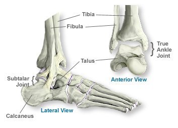
Three bones make up the ankle bone anatomy: Tibia (shin bone), Fibula (thin bone next to the shin bone), and the Talus (a bone of the foot that sits above the heel bone).
Bony protrusions can be seen and felt on the ankle. These bony bumps have their own names in ankle bone anatomy.
- Medial Malleolus: Bony bump on the inside of your ankle. The medial Malleolus is a part of the tibia’s base.
- Posterior Malleolus: Felt on the back of your ankle and is also a part of the base of the tibia.
- Lateral Malleolus: Bony protrusion felt on the outside of the ankle. The lateral Malleolus is the low end of the Fibula.
The ankle joint is not responsible for side-to-side movement of the foot. The tibiotablar joint gets replaced during ankle replacement surgery. The subtalar joint sits below the ankle joint and allows side-to-side motion.
Parts of the Ankle: Which ligaments make up the ankle joint?
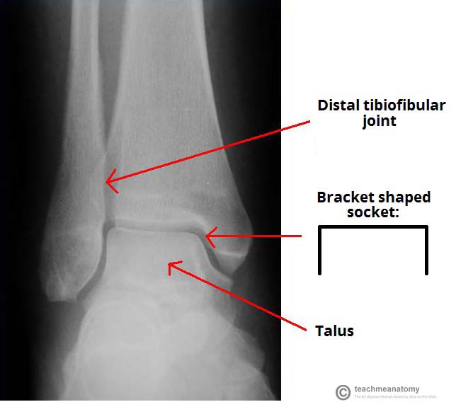
Along with the ankle bone anatomy, multiple ligaments surround both the ankle and Subtalar joints, which bind the bones of the leg to each other along with binding them to the bones of the foot. The Tibia and Fibula are connected by the Tibiofibular ligaments. This connection forms in the shape of a bracket and is covered in Hyaline cartilage. Hyaline cartilage is easily identifiable, known for its glass like appearance. It is pearly with a blue-gray tint in color and is slightly opaque.
Originating from the lateral Malleolus, the Lateral Ligament helps resist over-inversion (internal twisting) of the foot. The Lateral Ligament consists of three separate ligaments:
- Anterior Talofibular: Between the Lateral Malleolus and the lateral aspect of the Talus
- Posterior Talofibular: Between the Lateral Malleolus and the posterior aspect of the Talus
- Calcaneofibular: Between the Lateral Malleolus and the Calcaneus.
The Deltoid Ligament, or Medial Ligament, originates from the medial part of the distal Tibia and connects to the Medical Malleolus. The Medial Ligament consists of four ligaments which originate from the Malleolus and fan out to attach to the Talus, Calcaneus, and Navicular Bones. The Medial Ligament resists over-eversion (external twisting) of the foot.
Call 817-375-5200 to make an appointment with one of our foot and ankle specialists today!
F.A.Q.
What roles do the bones in the ankles play?
We can walk, run, jump, and engage in other activities thanks to the ankle bones’ support and stability for the foot and their capacity to move in the ankle joint.
How are fractures of the ankle bone handled?
The nature and degree of an ankle bone injury will determine the appropriate course of treatment. A cast or brace, physical therapy, or a visit to an orthopedic surgeon in Dallas may be needed to treat more severe injuries, while rest, ice, compression, and elevation can heal milder ones.
Can ankle bone injury lead to long-term issues?
Yes, if left untreated or if they happen frequently, injuries to the ankle bone can result in long-term issues like arthritis, persistent discomfort, and instability.
How can I avoid suffering an ankle bone injury?
It is crucial to use the proper footwear, warm up properly before physical activity, avoid uneven or unstable surfaces, undertake activities to strengthen the ankle and improve balance to prevent ankle bone injuries.